
The human foot is one of the most complex structures in the body. It supports balance, absorbs impact, adapts to uneven surfaces, and helps propel the body forward with every step. Although many people rarely think about foot anatomy until pain develops, understanding the structure of the foot and ankle can make it easier to recognize injuries, understand symptoms, and see how different conditions affect mobility.
One of the most common anatomy questions patients ask is: how many bones are in the foot? While the answer may seem straightforward, the structure of the foot is far more intricate than most people realize. Alongside these bones are joints, ligaments, tendons, muscles, and cartilage that work together continuously throughout daily movement.
How Many Bones Are in the Foot?
Each foot contains 26 bones, meaning there are 52 bones total in both feet. According to the American Academy of Orthopaedic Surgeons (AAOS), nearly one-quarter of all the bones in the human body are located in the feet.
Patients often wonder how many bones are in a foot because the foot must provide both flexibility and stability at the same time. These bones are divided into three major sections:
- Hindfoot
- Midfoot
- Forefoot
Each area contributes differently to balance, movement, and weight distribution.
The Hindfoot: The Foundation of Stability
The hindfoot forms the back portion of the foot and includes two major bones:
- Talus
- Calcaneus
The calcaneus, or heel bone, is the largest bone in the foot. It absorbs significant force during standing, walking, and running. Above it sits the talus, which connects the foot to the ankle joint and helps transfer body weight through the lower extremity.
Because this area handles repetitive impact throughout the day, hindfoot problems are commonly associated with heel pain, arthritis, tendon injuries, and fractures.
The Midfoot Supports the Arch
The midfoot contains five bones:
- Navicular
- Cuboid
- Three cuneiform bones
Together, these bones form the arch of the foot, which acts as a natural shock absorber during movement. Ligaments and tendons throughout this area help maintain stability while allowing the foot to adapt to different walking surfaces.
When patients ask how many bones of the foot are involved in supporting movement, the midfoot plays an especially important role because it helps distribute pressure evenly across the foot.
Problems affecting this area may contribute to:
- Arch pain
- Midfoot arthritis
- Tendon strain
- Flatfoot deformity
- Stress-related injuries
The Forefoot Helps Generate Motion
The forefoot includes:
- Five metatarsal bones
- Fourteen toe bones called phalanges
This section is heavily involved in balance and push-off during walking and running. The forefoot stabilizes body weight while the toes help generate forward movement.
Many common conditions develop in this area, including:
- Bunions
- Hammertoes
- Stress fractures
- Neuromas
- Arthritis in the toe joints
Understanding the name of the parts of the foot can help patients better identify where symptoms are occurring and communicate discomfort more clearly during evaluation.
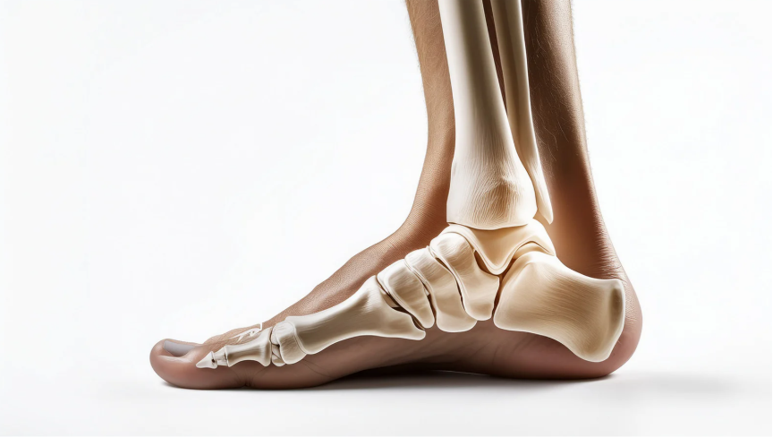
Understanding the Anatomy of the Ankle and Foot
The foot and ankle work together as a connected system. The anatomy of the ankle and foot includes bones, joints, tendons, ligaments, muscles, and cartilage that coordinate movement and stability.
The ankle joint itself is primarily formed by:
- Tibia
- Fibula
- Talus
These bones create the hinge-like structure that allows the foot to move upward and downward. Surrounding ligaments stabilize the ankle, while tendons help transfer force from muscles into motion.
One of the most important structures is the Achilles tendon, which connects the calf muscles to the heel bone and helps power walking and running.
Because the foot and ankle are closely connected, pain in one area can sometimes affect the other. This is why understanding overall foot and ankle anatomy can be helpful when evaluating persistent discomfort or instability.
Why Foot Anatomy Matters
Learning how many bones are in your feet is more than just an anatomy fact. The structures in the foot and ankle absorb substantial force every day while helping maintain posture, balance, and efficient movement.
During walking, the foot moves through a coordinated sequence:
- The heel absorbs impact
- The arch adapts to pressure
- The midfoot stabilizes the body
- The forefoot and toes push the body forward
This process occurs thousands of times daily. Even small problems involving alignment, joint mobility, or tendon function may gradually place excess stress on certain structures.
Understanding basic anatomy can also help explain why pain develops in different locations:
- Heel pain often involves the hindfoot
- Arch discomfort may involve the midfoot
- Pain near the ball of the foot commonly affects the forefoot or metatarsals
Many patients search for an ankle bones diagram when trying to understand injuries or symptoms more clearly. While anatomy resources can be helpful, foot and ankle pain is not always caused by bones alone. Tendons, ligaments, nerves, joints, and circulation can all contribute to discomfort.
If you are experiencing ongoing foot or ankle pain, swelling, instability, or difficulty walking, early evaluation may help identify the underlying cause before symptoms progress further. At Rocky Mountain Foot & Ankle, we provide comprehensive care for a wide range of lower extremity conditions. Patients in Murray can contact our team to schedule an appointment and learn more about treatment options tailored to their individual needs.